Mindfulness meditation is an important issue in this work. It fits nicely in the realm of reflectiveness because it is about opening the brain by rerouting the brain away from its ‘normal’ input. This stops the array of noise in the system, switching on (the cingulate cortex) our capacity to shape our narrative rather than be shaped by our old narratives.
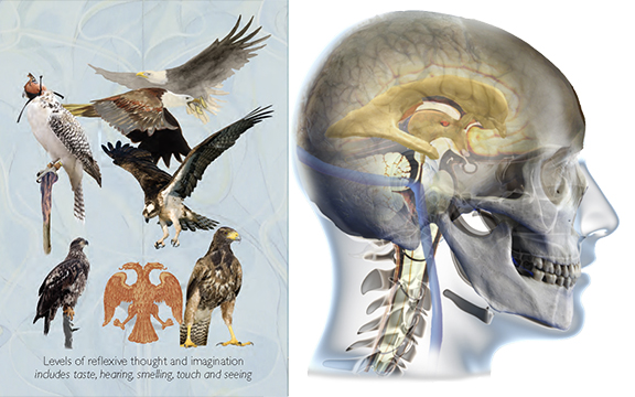
The varieties of reflectiveness and our ventricles as wings within our cerebrum
There are various ‘techniques’ that are in the general arena of mindfulness meditation. Some involve concentrating on breathing, and body parts, scanning for discomfort, unease or focusing on a particular organ (e.g. heart). Mindfulness meditation is about being attentive, taking our focus away from the input of extraneous noise. In this way, it helps us to make conscious decisions by helping to predict situations and avoid pain, injury, and death. It over-rides the subsystems which would deluge us into the noise of flight-fight, and the defensive pathway of both the hippocampus and amygdala.
The posterior cingular cortex is fully involved with reflectiveness or reflection. It involves cognitively aware attentiveness which involves deliberative reflection, the pulling back into focus, and a pin-point attention of or onto a stimulus. It also involves ruminative thought and a purposeful somatic awareness. The telling of narratives, stories, autobiographies, and experiences are all part of this part of the brain. In its subconscious functioning, it is loaded with data from social memes, social etiquettes, and other relevancies, and can automatically illustrate control of attention, irrelevant behavior, notice visual and somatosensory input or information that is useful and implement error correction, and pre-emptive action.
One of the useful protocols for mindfulness is to augment the brain’s coherence by subtly altering its inherent movement. In a state of coherence, the brain exhibits a particular minute but palpable movement (observed through human palpation). This inherent rhythm is postulated as a coherence or lack of coherence of many different rhythms, Entrainment allows for the various rhythms to come together to produce a coherent state which is picked up as a perceptible rhythm under the trained hands of an osteopath or similar therapist. When our bodies are in relative coherence then our physiological mechanisms appear to become more balanced and integral. In life, due to innumerable slight lesions from knocks, blows, jars, concussions, accidents, pathological or pathophysiological lesions, and original birth traumas, this normal movement may appear to be aberrant to the trained osteopathic physician, as these lesions alter the harmonics of all the mechanisms that (should be) in fluid rhythm.
The brain appears to move in three dimensions; vertically, sagittally, and rotatory. This is said to occur due to several factors.
-
The packing cells of the brain–the parenchymatous cells–imbibe cerebrospinal fluid–and are said to expand and contract as they fill and partially empty this fluid.
-
The ventricles–the centrally placed and paired structures that sit within the central core of the original notochord–filtrate cerebrospinal fluid via the choroid plexi which are housed in their roof. They secrete, fill, and expand with fluid, and then partially empty by allowing the fluid to drain through its dural barriers and various ‘doorways’ into various spaces within the meningeal coverings of the brain and spinal cord. This filling and emptying creates an expansion of the ventricles and their subsequent contraction. Because of the positioning of the brain within its dural housing, and bony structures, the brain rotates forwards and backwards.
-
The brain sits on a fibrous semi-elastic structure known as the falxes “cerebri and cerebellum” and a horizontally placed paired fascial floor called the tentoria. They create the internal struts of a balloon of dura which surrounds the brain and spinal cord. A rather delicate fulcrum exists where the falx and the tentoria meet. Any change of the internal dynamics creates a shift in this fulcrum, and because these fascia attach to the bones of the skull, they create a pull that helps the flexible sutures to move in a minute way which alters the whole skull’s shape as a structure.
The movement is such:
-
In its expansion phase, it moves backwards as if falling into the occipital (as if a cup) bone, as its horizontal structure (temporal lobes) expands anterolateral, and at the same time the vertical dimension shortens as the spinal cord rises upward and the brain flattens downward, together.
-
As the brain breathes out into extension, it involutes (pulls its lateral structures inward), as the whole structure moves anteriorly, and the vertical length elongates.
This mindfulness meditation has several variants.
We give them an initial graphic overview of the brain; the internal structures, the variable but relatively precise external markers for the structures, and the names of the various components and the movement ‘normally’ expected.
There are four stages.
Each position requires precise attention to promote a state of pause. This is elicited by placing one’s attention on a particular dynamic or place.
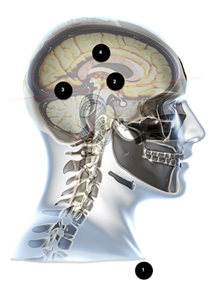
Each part is labelled
-
Breath
-
Midbrain / 3rd ventricle
-
Sutherland’s fulcrum – the approximate meeting point of the four sickle-shaped membranes of the internal struts of the dura mater.
-
Paired and central ventricles
1–Mindfulness through Breath Technique
In this meditation, they are asked to watch their breath. We ask them to be present with the space or pause elicited prior to the next breath. That is, they observe their in-breath and out-breath and notice that they can create, cause, or notice that often, but not always, there will be a pause between the next in-breath. They ought to watch passively without interference with their breathing cycle. Initially, they may notice an irregularity or some anomaly in breathing, but slowly, with their attention on breathing most people can easily achieve a gentle rhythmic natural cycle. There is a space, a point, an interval between breaths that is the focus of this type of meditation. If they concentrate on this moment each time, their awareness and attentiveness are then focused (integrity) in that they have pinpointed a particular moment. This refocuses the brain into non-doing, apart from watching for that moment. It helps to censor the continual noise of thought.
For many people, this gentle meditative process brings them into a natural quiet and ease. Mindfulness at this level is not surrender, but it opens the possibility for the brain to be open, receptive, and at ease so that in surrender it can act as an interpreter for new information.
2–Mindfulness through attention on the midbrain/hypothalamus
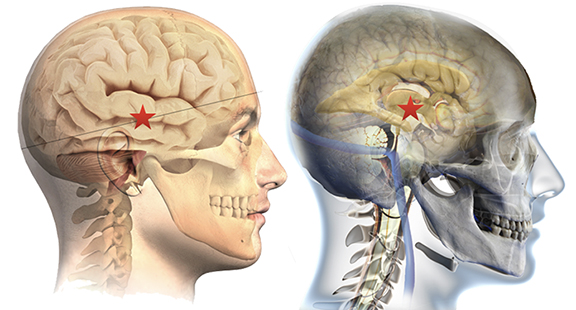
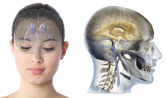
Location of hypothalamus
The hypothalamus can be seen as the centre of the brain. It exists at the top of the central neural core, which houses important nuclei that act like a biochemical laboratory. It is the central foci for the vegetal system because it monitors the chemistry, and then initiates global changes through the pituitary and pineal glands.
You can locate the hypothalamus which as a structure, is the floor of the 3rd ventricle. This is a small rhomboidal area filled with cerebral spinal fluid, and its lateral walls are made up of an important relay and translator nuclei–the thalamus–for sensory input, executive parts of the cortex, and the limbic system. The person would simply locate the approximate midline structure, from just above their frontal eminences to the back of the head where the person can feel the corrugations across the bone called the occiput. Laterally, the hypothalamus is approximately above and slightly forward of the ear–similar to the picture. The participant puts their attention on this location inside their brain and simply keeps it there for as long as they can. If their attention slips, then they simply pull it back.
-
This exercise of quietness helps to reset the biochemistry of the body.
-
It helps to reset the pituitary and pineal as both extrude through recesses at the front (pituitary) and back (pineal) of the 3rd ventricle.
-
It has a relationship with the central core circuit of the meridian system.
3–Mindfulness through attention on the cerebral vein on the meeting point of the four internal dural struts
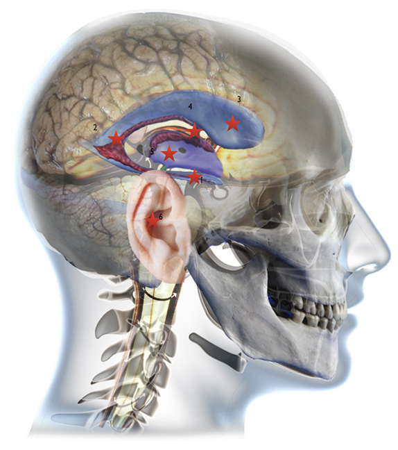
Technical anatomy
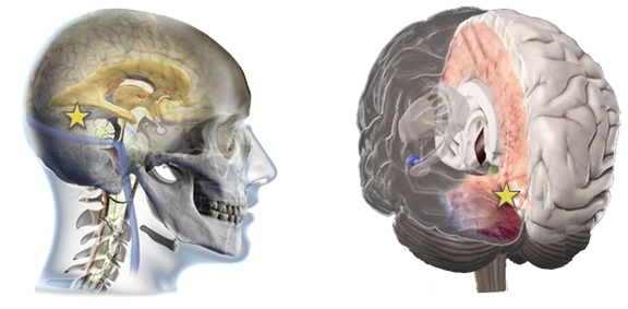
The meeting point at the straight sinus
The brain is divided by four infoldings of the two layers of the dura. One, the outer layer, attaches to the periosteum or underneath a layer of the bone, the other splits and reduplicates creating folds that divide the two cerebral hemispheres, and create a floor for the hindbrain, and its doubled-up partner, the roof for the cerebellum. A small vertical continuation of the dura creates a partial division of both cerebellum. Where the four meet there is a space within which the great cerebral vein (the Great vein of Galen) runs through helping to empty the internal structures of deoxygenated blood. Where each fold originates in the periosteal layer, a blood sinus helps to empty the main cerebral blood flow from the interior of the brain. The sinus is constructed by the bifurcation of the dura from the periosteal layer.
Osteopaths consider this meeting point of the four sickle-shaped membranes as a fulcrum. This fulcrum appears to move as the brain and dural envelope reciprocate very slightly as the cranium breathes in and out, as the CSF fills and partially empties within the internal cerebral spinal flow–through the ventricles–and the sinus regulation of fluid.
This meditation asks the participant to place their attention upon the approximate position of this fulcrum. It can be sensed as if it were about 15 cm inwards, within the brain, from the central corrugated ridging found on the back of our occiputs. It is slightly sloped, going up towards the centre. If we were to draw a line from the glabella–between the eyebrows–and the corrugations you would have an approximate sense of the incline. The spot actually is moving dependent upon the relative phase of the cranial movement, but suffice it to say that if you place your attention upon it, then the movement stops as the whole mechanism finds a balanced place, re-organizing itself. If your mediation is 15 or twenty minutes, this will suffice to reset this fascial reciprocating fulcrum which will help balance all the other fascia in the body.
4–Mindfulness through attention to the ventricles and their movement
In their meditation, they are asked to envisage and watch for the movement of the ventricles. They impose their thoughts upon the structure. What we know is that thought is followed by movement, as energy follows thought or mind. We get the participants out of their reflexive brain into quietness, but at the same time, we get them to notice the movement–real or imagined! They should imagine and sense the rotation of the occiput with the following pull of the frontal bone. They ought to sense the expansion or not, whether equal, distorted, or absent, of the lateral components. They ought to note the shortening of the spinal core in flexion; or not.
Ventricles on view
They then should note the gentle 3-second rhythm where the brain changes the filling phase and begins to partially empty its (cerebrospinal) fluid, which will circulate around the brain and down the meningeal layers of the spinal cord out into the neurolemma of the peripheral nervous system. As it does so, they will try to sense and become aware of the frontal mass (bone) moving ever-so-slightly forward, as the occiput rotates forwards and upwards, as if it were to cup the brain. The lateral components pull inward. This feels as if the mass between the ears shrinks inward. The opposite movement is when the occiput drops and rotates backwards, pulling on the frontal, whilst the top of the head flattens, and the side bones–the temporals–rotate forwards and slightly outwards, expanding the width of the brain.
4b – Swimming through the ventricles
We get the participants to envisage the internal structure of the ventricles and for them to travel through them as if swimming. We get each person to metaphorically dive in from the lateral limbs of the lateral ventricles and to travel through the water-filled space, down through open doorways, and into submerged spaces.
What does this do?
It provides an attentiveness to be present in a focused manner. This stops the monkey mind (limbic and amygdala noise-driven thoughts) and gets our attention. It initiates our cingulate cortex.
The swim through the ventricles also provides an exercise in flushing and flexing our internal structure. The roof of the paired lateral ventricles is made up of the hippocampus–a very important animal-vegetal part of our brain that processes emotion, and memory, particularly short-term or present time. As we swim through these paired structures the structures appear to flex and change via our attention. Sometimes as we ‘swim’ through these structures we find ourselves ‘somewhere’ else, as if thrown out of the structure. This is normal. It is as if we have encountered a force within the structure that is bigger than us, and we exit the area in our own flight-flight reaction. We ask the participants to come back until they are able to swim through the structure.
On other occasions they will be unable to swim; as if encountering a wall or some force which prevents their forward motion. They simply stay there until the fluid gives way and they can again swim.
Sometimes they won’t be able to go through the various doorways or foramen. It is as if the doors are shut. Again they simply wait until they are allowed through.
We ask them to go down through the central canal (of Majendie) and into the fourth ventricle. They can exit through the back of this small tent-like structure through the two openings which flush out some of the fluid into the cerebellar cistern under the cerebellum at the back of the brain. They can then exit the brain from there.
-
Dive in, laterally and simultaneously both sides of the brain, into the lateral ventricles (just the infant of the ears and slightly above them) and swim backwards towards #2.
-
This is deeper into the cerebrum and is both a chamber and a corner, which you turn around and swim uphill, if you are lying down, or forwards and initially slightly upwards if you sitting.
-
Go to the bulbous nose of the ventricle, and look backwards and down; you will find that both ventricles are paired and that there is a small opening in the floor.
-
Swim through these two openings into a common single, central tight space; the third ventricle. (At its front bottom end is a recess for the pituitary, and at the rear end, a recess for the pineal–both of which protrude outwards as distinct structural entities.) The third ventricle’s floor makes up the nuclei structures of the hypothalamus, and the vertical walls are relay substations made up of much of the connections going through and from your brain–the thalami.
-
In the middle floor is a little opening, the size of a straw diameter, and a long tube that allows you to slide down into the fourth ventricle.
-
The fourth ventricle is directly behind the cerebellum.
-
In its wings are two sleeves or openings that allow you to swim out of the tent-like cave and into a waterbed under the cerebellum.
-
From there you can gently re-emerge back into your normal world.
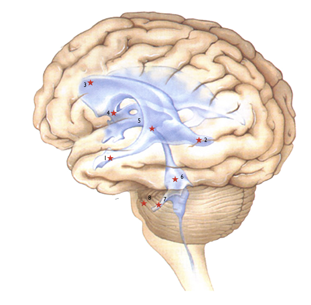
Numbers relate to the various structures you will meet